A routine eye examination prevented a 57-year-old Filipina resident of Sharjah from going blind for the rest of her life. The expat had been suffering from vision loss and heaviness around her right eye for almost a month when she consulted an ophthalmologist. She was then referred to an Oculoplasty and Orbit Surgery specialist, who discovered a large tumor in the orbit of her eye and recommended further examination. A massive tumor, measuring 35×30 mm—larger than the patient’s 24 mm eyeball—was successfully removed following a strenuous, hour-long, complex surgical procedure.
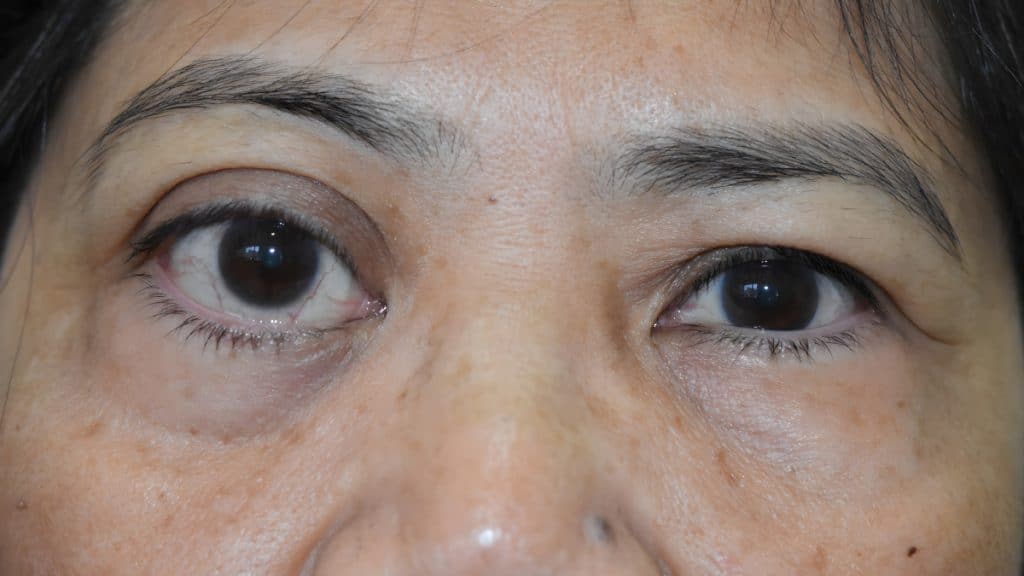
The astute ophthalmologist noticed that the patient’s right eye protruded more than her left during the eye checkup. The patient explained that it was a slowly progressive condition that had been evident since her childhood. Following the ophthalmologist’s timely diagnosis, an MRI and CT scan were conducted, revealing a well-defined oval intraconal lesion in the right orbit, indicating the likelihood of a cavernous hemangioma (a vascular tumor). The patient underwent a highly sophisticated, micro-invasive surgery—believed to be the first of its kind at the hospital—to remove the tumor from behind her eye.
Cavernous hemangioma is a benign vascular tumor commonly discovered in middle age. It is one of the most frequent benign tumors found in the orbit of the eye. While slow-growing, when situated near critical areas like the optic nerve, it can cause vision loss or blindness over time. In this case, the tumor was compressing the optic nerve, threatening the patient’s vision.
Dr. Fairooz P.M., the Oculoplastic Surgeon who led the surgical team, stated, “This was a very risky case, as the tumor was compressing the optic nerve and causing vision loss. The surgical procedure involved protecting the optic nerve to regain vision. This meticulous surgery, which required a high level of precision, was crucial in preventing further vision deterioration.”
Before the surgery, the tumor caused the patient’s eye to protrude forward, threatening her vision by compressing the optic nerve. A micro-incision internal orbital surgical approach was adopted. According to Dr. Fairooz, the benign tumor, which measured 35×30 mm, was larger than the patient’s 24 mm eyeball. Though the tumor had not spread to other areas of the eye, it continued to grow, compressing the optic nerve and causing compressive optic neuropathy.
Highlighting the critical nature of the case, Dr. Fairooz said, “Cavernous hemangioma is one of the most common orbital tumors, accounting for 20 percent of all orbital masses. While these tumors are usually painless and slow-growing, they can pose serious challenges when located near crucial structures like the optic nerve, potentially threatening vision.”
The diagnosis and management of orbital tumors, particularly those affecting visual function, pose significant challenges for surgeons. In such cases, timely and precise intervention is essential to prevent serious complications, such as vision loss.
Following the complex procedure, the patient’s vision was completely restored. Reflecting on the outcome, Dr. Fairooz expressed gratitude to the entire team for their combined efforts, which made the treatment successful. She added, “We are very pleased that the patient responded well to the surgery and has fully recovered. The bulging of the eye was actually the symptom that indicated the tumor’s presence, making it a crucial component of the diagnosis. The pressure on the optic nerve was more harmful and threatened the patient’s vision.”
Dr. Fairooz continued, “We believe this is the first time such a procedure has been carried out by the Oculoplasty and Orbit Surgery department at the hospital, and we are delighted with the patient’s recovery.”
The multidisciplinary approach taken by the hospital was key to the success of this complex procedure. The collaborative efforts of the oculoplastic surgeon and radiologists resulted in an accurate diagnosis. A well-executed treatment plan, which utilized advanced imaging techniques, cutting-edge surgical methods, and skilled operation theatre staff, was instrumental in achieving a positive outcome for the patient, showcasing the hospital’s expertise in managing such challenging cases.